Inferior orbital fissure
Gap between bones of the human eye socket
| Inferior orbital fissure | |
|---|---|
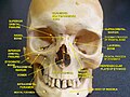
 The skull from the front. (Label for inferior orbital fissure is at center right.) | |
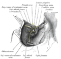
 1 Foramen ethmoidale, 2 Canalis opticus, 3 Fissura orbitalis superior, 4 Fossa sacci lacrimalis, 5 Sulcus infraorbitalis, 6 Fissura orbitalis inferior, 7 Foramen infraorbitale | |
| Details | |
| Identifiers | |
| Latin | fissura orbitalis inferior |
| TA98 | A02.1.00.084 |
| TA2 | 489 |
| FMA | 54802 |
| Anatomical terminology [edit on Wikidata] | |
The inferior orbital fissure is a gap between the greater wing of sphenoid bone, and the maxilla. It connects the orbit (anteriorly) with the infratemporal fossa and pterygopalatine fossa (posteriorly).[1]: 397
Anatomy
The medial end of the inferior orbital fissure diverges laterally from the medial end of the superior orbital fissure. It is situated between the lateral wall of the orbit and the floor of the orbit.[1]: 397
Contents
The fissure gives passage to multiple structures, including:
- Infraorbital nerve,[1]: 402 artery[1]: 364 and vein[citation needed]
- Inferior ophthalmic vein[1]: 364, 403
- Zygomatic nerve[1]: 496
- Orbital branches of the pharyngeal nerve[1]: 370
- Maxillary nerve
Additional images
-
 Left infratemporal fossa.
 -
 Horizontal section of nasal and orbital cavities.
 -
 Dissection showing origins of right ocular muscles, and nerves entering by the superior orbital fissure.
 -
 Inferior orbital fissure.


See also
References
![]() This article incorporates text in the public domain from page 189 of the 20th edition of Gray's Anatomy (1918)
This article incorporates text in the public domain from page 189 of the 20th edition of Gray's Anatomy (1918)
- ^ a b c d e f g Sinnatamby, Chummy S. (2011). Last's Anatomy (12th ed.). ISBN 978-0-7295-3752-0.
External links
Wikimedia Commons has media related to Inferior orbital fissure.
- lesson3 at The Anatomy Lesson by Wesley Norman (Georgetown University) (orbitforamina) (#3)
- "Anatomy diagram: 34256.000-1". Roche Lexicon - illustrated navigator. Elsevier. Archived from the original on 2012-12-27.
- "Anatomy diagram: 34257.000-1". Roche Lexicon - illustrated navigator. Elsevier. Archived from the original on 2012-07-22.
- v
- t
- e
Foramina of the skull (and canals, fissures, meatus, and hiatus)
| to Orbit: | |
|---|---|
| to Nasal cavity: |
|
| to Orbit: |
|
|---|---|
| to Pterygopalatine fossa: |
|
| to Infratemporal fossa: |
|
| other: |
- internal auditory meatus/facial canal/stylomastoid foramen (CN-VII,VIII)
- jugular foramen (CN-IX,X,XI)
- foramen magnum (CN-XI)
- hypoglossal canal (CN-XII)
- condylar canal
- mastoid foramen
| to Nasal cavity: | |
|---|---|
| to face: | |
| to Pterygopalatine fossa: |
|
| other: |
|
| to Nasal cavity: | |
|---|---|
| to Oral cavity: | |
| to Infratemporal fossa: | |
| to Nasopharynx: |
| to oral cavity: | |
|---|---|
| to nasal cavity: |
|
Portal:
 Anatomy

| Authority control databases |
|
|---|


